IDEXX Reference Laboratories
DISCOVER MORE
IDEXX clinical pathology services
Faster turnaround means faster treatment.
IDEXX cytology services are designed to provide accurate, timely results when you need them to make confident treatment decisions.
- Largest global network of veterinary clinical pathologists, offering their expertise to support you in making clinical decisions.
- Integrated, comprehensive reports with images.
- Comprehensive cytology educational resources .
What's new.
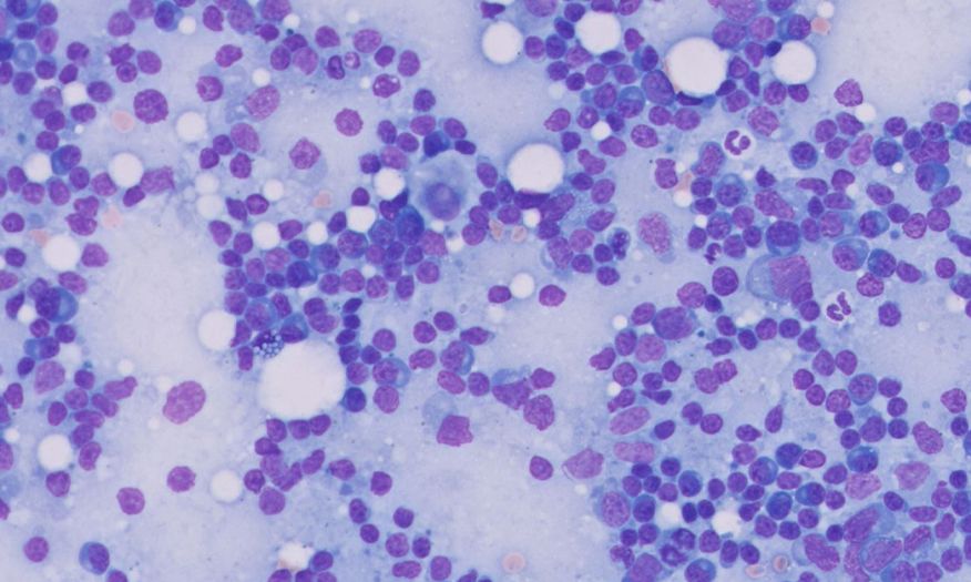
Reference laboratory submissions—Lymph Node Cytology services
- All lymph node submissions will be processed under the following new test codes:
- 609A: Samples from multiple lymph nodes
- 6091A: Lymph node and one mass
- 6092A: Lymph node and two masses
- New test codes to ensure the most appropriate processing of your specimen and allow for the appropriate larger number of slides associated with these cases.
- Includes cytology with microscopic description for one mass or two masses, as appropriate, and multiple lymph nodes.
Comprehensive testing services to cover even your most time-sensitive cases.
If your case is clinically urgent, please make a note on the submission form and we will prioritise wherever possible.
1-2 days after day of receipt into the laboratory (Mon-Fri)
- Quick, reliable turnaround for your routine cases
- Personalised guidance from our team of expert pathologists
Test codes
CYT: 1 site/lesion
CYT2: 2 sites/lesions
See the online Directory of Tests and Services at vetconnectplus.com.au
for test codes for additional sites/lesions.
1-2 days after day of receipt into the laboratory (Mon-Fri)
- Personalised guidance from our team of expert pathologists
Test codes
609A: Lymph Node Cytology
6091A: Lymph Node Cytology with 1 Mass/Lesion
See the online Directory of Tests and Services at vetconnectplus.com.au
for test codes for additional sites/lesions.
1-2 days after day of receipt into the laboratory (Mon-Fri)
- A full summarised clinical history and a concurrent CBC result and blood smear submission (within 24–48 hours of marrow collection) is required for complete interpretation and clinical commentary.
Test code BME
1-2 days after day of receipt into the laboratory (Mon-Fri)
Test codes
BFLA: 1 site
BFLA2: 2 sites
See the online Directory of Tests and Services at vetconnectplus.com.au
for test codes for additional sites/lesions.
1-2 days after day of receipt into the laboratory (Mon-Fri)
Test codes
CJO: 1 site
CJO2: 2 sites
See the online Directory of Tests and Services at vetconnectplus.com.au
for test codes for additional sites/lesions.
1-2 days after day of receipt into the laboratory (Mon-Fri)
Test code CSFLA
Note: Please specify site of collection as either the cerebellomedullary cistern or lumbar cistern when collecting CSF.
1-2 days after day of receipt into the laboratory (Mon-Fri)
Test code WFLA
Note: Please specify whether sample was collected as a BAL or TW.
Featured case
Swollen lymph node diagnosis.
A 14-year-old male Yorkshire terrier named Charlie presented with a firm swelling under the jaw and decreased appetite. Specimens were collected for an IDEXX CBC, serum chemistry including an IDEXX SDMA Test, total T4, and complete urinalysis. Fine needle aspirates were collected from the prescapular, axillary, inguinal and popliteal lymph nodes, as well as the left mandibular lymph node. Slides were prepared from the aspirates, and all specimens were submitted to IDEXX Reference Laboratories. Diagnostics revealed mild nonregenerative anaemia, mild neutrophilia, monocytosis, eosinopaenia, increased SDMA, ALP, amylase and lipase levels, and decreased albumin. Charlie was diagnosed with multicentric large cell lymphoma, an aggressive form of cancer. The client declined further diagnostic testing and opted for palliative treatment with prednisone.

Best practices on how to make, prepare, and pick the best slides for submission.
Always remember to:
- Label each slide with the patient name, date, source, and preparation technique.
- You are welcome to submit prestained slides for review, as well as unstained slides in-clinic to check the quality of the preparation, cellularity, and cell preservation.
Read more about how to prepare slides
Accurate and timely cytology results start with good quality slides
Fine needle aspiration and nonaspiration
- Use for skin/subcutaneous masses, lymph nodes and internal organs.
- Select the technique best suited for the tissue to avoid cell lysis or excessive blood.
- Use 22-gauge needles, which work well for most tissue types.
Blood films
- Use fresh, well-mixed anticoagulated blood to avoid specimen deterioration.
- Maintain an approximate 30° angle between the spreader and specimen slide throughout.
- Spread the specimen in one smooth, steady motion.
Line smears
If submitting fluids for cytologic evaluation, send one EDTA tube as well as a plain, sterile tube particularly if culture may be requested.
If possible, preparation of a direct smear or a line smear may assist with optimal sample preservation.
- Use for body cavity effusions, bronchoalveolar lavage (BAL)/tracheal, lavage, and joint fluid.
- Include a direct smear; use an additional line smear for specimens with low cellularity or suspected infectious agents. If needed, use a squash prep for flocculent material.
- Submit a buffy coat and a direct film for specimens with large blood content.
Urine sediment
- Use 5 ml of fresh, well-mixed, centrifuged urine (set centrifuge to urine/400 x g).
- Maintain an approximate 30-40 angle between the spreader and specimen slide, using the resuspended pellet.
- Spread the specimen halfway down the slide; stop abruptly to create a line.
- Specify on the submission that the sample is a sediment preparation.
- Send the urine sample also for the lab to make additional preparations.
Always remember to:
- Follow the recommended stain times.
- Check slides to see if additional staining is required.
- Replace stains as needed to prevent stain precipitate.
- Use separate stain sets for "clean" cytology (e.g., mass aspirates) and “dirty” cytology (e.g., ear and skin cytology).
Step 1. Dip in fixative
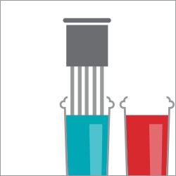
Step 2. Dip in red stain
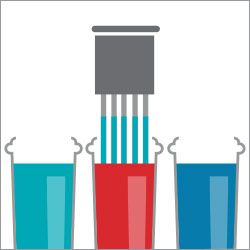
Step 3. Dip in blue stain
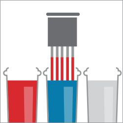
Step 4. Dip in water
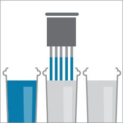
Step 5. Dip in water
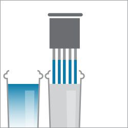
For in-depth instruction and CE credit at the IDEXX Learning Centre:
Preparing slides reference guide
IDEXX Learning Centre education
Choosing the Best Slides to Submit for Pathology Review
Getting the Most Out of Your Cytology Pathology Submissions
The Differences Between Histology and Cytology
Cytology Techniques
FAQs
- Leverage resources through the IDEXX Learning Centre to help your staff with optimal slide preparation to ensure they are able to prepare and select the best slides. This will not only save you time but also ensure we can provide you with timely and accurate results.
- Providing more clinical details on the request form will also assist the pathologist in providing the most accurate diagnosis for the patient and the pet owner. Please see the Pathology Submission Guidelines for complete details.
- We encourage practices to reach out to the pathologist to discuss their cases. The pathologist’s direct contact information is provided on the result report.
Pathology consultation services are complimentary for IDEXX customers, even before a specimen is submitted. We encourage practices to reach out to the pathologist to discuss their cases. The pathologist’s direct contact information is provided on the result report. For questions regarding pre-submission pathology support, contact IDEXX customer support at 1 300 44 33 99 .
Images are captured by the pathologist using a scope camera for glass slides and digital image capture software on digitised slides. IDEXX provides an image in both clinical and anatomic pathology reports at no additional charge to the customer exclusively via VetConnect PLUS , including the VetConnect PLUS mobile app, for a more comprehensive report. Reports with images in VetConnect PLUS are printable and can be shared.
Support
Documents and other resources
Access IDEXX Reference Laboratories
guidelines
, IDEXX Online Orders
, and educational resources
.
Directory of Tests and Services
Browse our tests and services to see a comprehensive list of offerings from IDEXX Reference Laboratories and save them to VetConnect PLUS so you can easily access them anytime.
IDEXX Reference Laboratories Support
We support your practice with our customer support, technical support, and medical consulting services team, including our board-certified veterinary specialists.
Call us at 1 300 44 33 99 .
Learn more about a specific product or service.
A representative will help you every step of the way.
IDEXX Reference Laboratories
DISCOVER MORE
Note: There may be times where weather or operational considerations cause delays in providing test results. When this happens, we will inform you as quickly as possible with the most complete information available.
