IDEXX Newsletter
Alice Gee, our Veterinary Laboratory Technician takes us through the steps on how to make a good blood smear.
Making a good quality blood smear is essential, and may take some practice. To help, here is a 4-step guide,
Step 1: Ensure your spreader slide is clean, and you have all the necessary equipment ready.
We recommend a small amount of saline in a container to rinse your spreader slide between smears. Making smears from EDTA blood is preferred, but citrate and heparin are also acceptable. If you do use citrate or heparin, please be sure to note this on the smear or on your submission form.
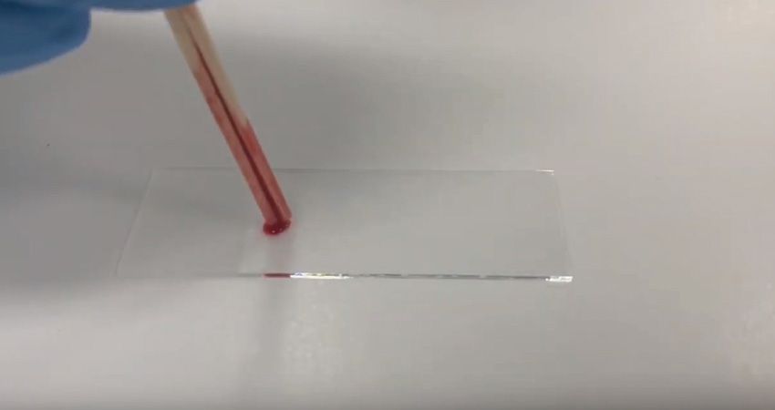
Step 2: Thoroughly mix your blood, then fill a capillary tube. Using the capillary tube place a small amount of blood about 1cm from the end of slide.
Try to minimise or avoid getting bubbles in the blood when putting it on the slide. Check to see if your slide is clean too, as any dirt or dust will be drawn through your smear.
Step 3: Place your spreader on the slide at a 45o angle, then pull the spreader back into the blood. The blood will then traverse along the edge of the spreader – once it has reached the very edge, push the spreader forward in a rapid, smooth motion.
When making smears on anaemic or thick bloods, it may be necessary to adjust the angle and speed. IFor anaemic samples we recommend increasing the angle between the spreader and slide. You can also try pushing the spreader faster. The opposite applies to thicker bloods which may not spread very far at all. Decrease the angle for these bloods, and push the spreader a bit slower.
Step 4: Let your smear dry, then send it with the blood sample and request form to your nearest IDEXX Laboratory. Ideally a blood smear should be sent with every CBC submission.
Let the smear air dry gently – it’s best not to speed up the process with a hair dryer or putting it against something hot. If possible, send the EDTA blood with an unstained smear. You can also make an extra smear to stain and examine yourself! A good quality smear has a clear, feathery end to it, where you can see individual cells in a single layer under the microscope.

